Because this piece does not have an abstract, we have provided for your benefit the first 3 sentences of the full text.
Have you ever wondered if and how often an electrocardiogram (EKG) should be obtained to monitor the QTc? Have you been unclear about which medical and metabolic conditions and medications can prolong the QTc? If you have, then this case of an elderly man with symptoms referable to multiple organ systems and treatment that involved several agents with QTc-prolonging side effects should prove helpful.
Recommendations for QTc Monitoring:
Rational or Arbitrary?
LESSONS LEARNED AT THE INTERFACE
OF MEDICINE AND PSYCHIATRY
The Psychiatric Consultation Service at Massachusetts General Hospital (MGH) sees medical and surgical inpatients with comorbid psychiatric symptoms and conditions. During their twice-weekly rounds, Dr Stern and other members of the Consultation Service discuss diagnosis and management of hospitalized patients with complex medical or surgical problems who also demonstrate psychiatric symptoms or conditions. These discussions have given rise to rounds reports that will prove useful for clinicians practicing at the interface of medicine and psychiatry.
Prim Care Companion CNS Disord 2018;20(5):18f02327
To cite: Benjamin LN, Elshafey S, Kistler EA, et al. Recommendations for QTc monitoring: rational or arbitrary? Prim Care Companion CNS Disord. 2018;20(5):18f02327.
To share: https://doi.org/10.4088/PCC.18f02327
© Copyright 2018 Physicians Postgraduate Press, Inc.
aDepartment of Medicine, Massachusetts General Hospital, Boston, Massachusetts
bHarvard Medical School, Boston, Massachusetts
cDepartment of Psychiatry, Massachusetts General Hospital, Boston, Massachusetts
‡Authors contributed equally to this article.
*Corresponding author: Theodore A. Stern, MD, Department of Psychiatry, Massachusetts General Hospital, Fruit St WRN 605, Boston, MA 02114 ([email protected]).
Have you ever wondered if and how often an electrocardiogram (EKG) should be obtained to monitor the QTc? Have you been unclear about which medical and metabolic conditions and medications can prolong the QTc? If you have, then this case of an elderly man with symptoms referable to multiple organ systems and treatment that involved several agents with QTc-prolonging side effects should prove helpful. We use this case as a stimulus for discussion, review the risk-benefit analysis for medication administration and monitoring, and discuss the rationale for daily QTc monitoring.
CASE VIGNETTE
Mr A, a 68-year-old man with a history of smoking, chronic obstructive pulmonary disease (COPD), heart failure (with a preserved ejection fraction), chronic lower back pain, posttraumatic stress disorder (PTSD), depression, and insomnia, was admitted to the hospital with fever, acute-onset dyspnea, cough, and lower-extremity edema. He had been in his usual state of health until 3 days earlier when he developed bilateral lower-extremity edema. The following day, he felt feverish and increasingly dyspneic and was coughing. His dyspnea worsened over the next 2 days, and he came to the emergency department (ED) for evaluation.
On arrival to the ED, he was noted to be afebrile; however, his heart rate was 152 bpm, he was tachypneic, and his blood pressure was 85/56 mm Hg. The physical examination was notable for respiratory distress, diffuse wheezing, and mild symmetric bilateral lower-extremity edema. Mr A was treated with 2 L of crystalloid fluid, and he was started on bilevel positive airway pressure. Laboratory evaluation revealed a neutrophil-predominant leukocytosis to 18.9 cells/mm3, and his chest x-ray showed a patchy opacity in the left lung base. His initial EKG revealed sinus tachycardia with a QTc of 441 ms. Mr A received ipratropium-albuterol nebulizers, intravenous (IV) methylprednisolone, and IV magnesium, as well as vancomycin and cefepime for a presumed COPD exacerbation that was triggered by pneumonia. His antibiotic regimen was quickly modified to levofloxacin (750 mg daily) for treatment of community-acquired pneumonia.
Mr A took methadone (20 mg 4 times daily), tizanidine (8 mg twice daily), and pregabalin (200 mg 3 times daily) for chronic low back pain; mirtazapine (45 mg nightly) and quetiapine (250 mg daily) for persistent depression; and trazodone (200 mg nightly), prazosin (2 mg), and zolpidem (5 mg intermittently) for severe insomnia secondary to posttraumatic stress disorder (PTSD).
Given his borderline elevated QTc (441 ms), use of multiple medications known to affect the QTc, and the recent addition of levofloxacin (750 mg daily) to his medication regimen, the medical team obtained a daily EKG to monitor for the development of QTc prolongation.
DISCUSSION
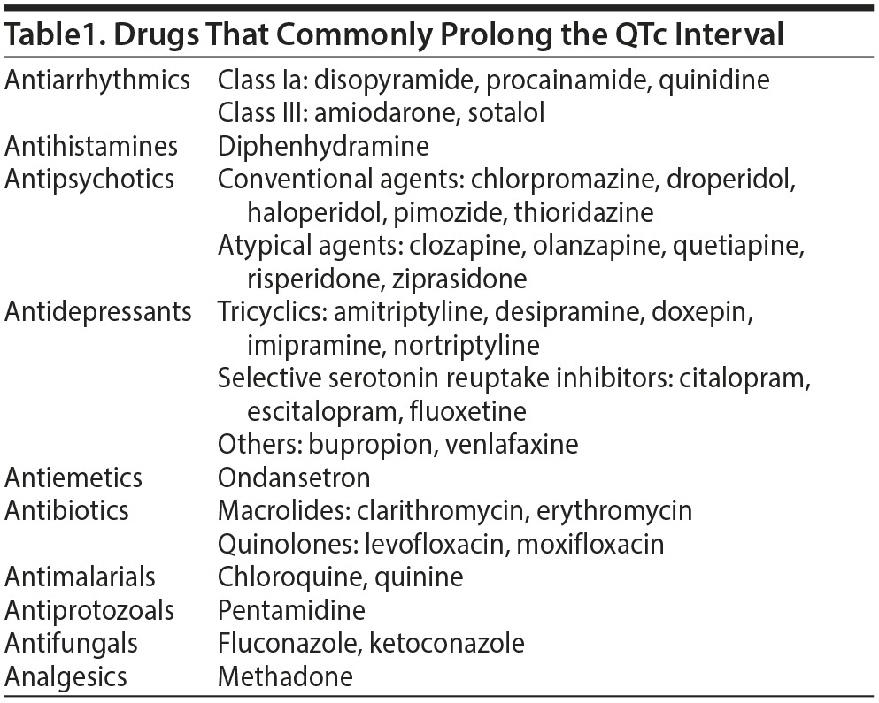
Across the United States, patients who require hospital-level care are typically treated with multiple medications, and many of these agents prolong the QTc interval, directly or indirectly, via drug-drug interactions (Table 1). Unfortunately, when the QTc increases to > 440 ms, the risk of R on T phenomena and lethal arrhythmias (eg, torsades de pointes [TdP], ventricular tachycardia, ventricular fibrillation) also increases. Clinicians’ fears of inducing QTc prolongation and lethal arrhythmias lead to frequent monitoring (eg, via daily EKGs or an EKG after each dose increase of a medication associated with QTc prolongation). These monitoring strategies are accompanied by costs (eg, of time, effort, and money) and are often associated with undue concern by patients, providers, and systems of care (eg, concern over liability if diligent monitoring does not occur).
What Is the QTc and What Prolongs It?
The QT interval is the time between the start of ventricular depolarization and the end of ventricular repolarization. On a standard 12-lead EKG, the QT interval is measured from the junction of the isoelectric baseline and first negative deflection in the QRS complex to the end of the T wave, specifically, to the point where the tangent with maximum slope (second half of the T wave) intersects with the isoelectric line. The QT interval is rate-dependent, and reference values are normalized to heart rate, generating the QTc or the QT interval divided by the square root of the R-R interval. Measurements of QTc are imperfect and depend on the lead being analyzed, with leads II, V5, and V6 yielding the most accurate measurements. Furthermore, the QTc varies from moment to moment and is dependent on several factors including, but not limited to, autonomic tone, position, and volume status. The QT interval can also be affected by pharmacotherapies1; it is classically prolonged by class I and III antiarrhythmics. Numerous commonly used classes of medications (eg, antipsychotics, antidepressants, analgesics, antibiotics) have also been implicated in QT prolongation.2,3
Medication-mediated QT prolongation is of clinical importance because it is associated with the development of TdP, a life-threatening polymorphic ventricular tachycardia. Importantly, however, while QT prolongation can precipitate TdP, it is not sufficient by itself. The substrate for this arrhythmia instead relies on the transmural dispersion of repolarization, meaning that the time for ventricular repolarization varies from endocardium to epicardium, allowing for reentrant excitation. This dynamic is best exemplified by the class III antiarrhythmic amiodarone, which prolongs the QTc on the surface EKG but still ultimately suppresses arrhythmias by standardizing repolarization times across the cardiac wall and, thereby, decreasing the risk of reentrance and the generation of TdP.2
What Is the Prevalence of QTc Prolongation?
Much of the data on the prevalence of QTc prolongation among inpatients comes from small, single-center studies4–6 with significant limitations. For those admitted to medical services, some degree of QTc prolongation appears to be common. One prospective study4 found that 24% of critically ill intensive care unit patients had a QTc > 500 ms. A small retrospective study5 of elderly patients admitted to an acute geriatrics service noted that 27% had QTc prolongation (defined as ≥ 470 ms in women and ≥ 450 ms in men). Similar rates of QTc prolongation were seen in a prospective observational study of 279 patients in the ED; 34.1% of them had a prolonged QTc interval (defined as ≥ 460 ms in women and ≥ 450 ms in men).6 The prevalence may be even higher in those with an underlying cardiac disease, as demonstrated by a prospective observational study7 of cardiac patients that showed that 44.6% had some degree of QT prolongation and 17.3% had a QTc > 500 ms. Interestingly, in a large retrospective study8 of 41,649 hospital admissions, only 293 patients (0.7%) had a QTc > 500 ms; this finding most likely reflects that this study was not limited to medical patients (who are more likely to have underlying comorbidities or coadministered medications that contribute to a prolonged QTc).
How Frequently Are QTc-Prolonging
Medications Coadministered?
Medical inpatients over the age of 75 years receive a mean of 7.5 drugs each day; thus, the risk of QTc prolongation is increased in this group, as this population is at high risk for delirium and treatment with antipsychotics that prolong the QTc interval.9 Among inpatients, prescription of antiarrhythmics, antiemetics, antibiotics, analgesics (eg, methadone), or antipsychotics—especially when hypomagnesemic, hypokalemic, and bradycardic—create a “perfect storm.” One cross-sectional study10 showed that 0.9% of psychiatric inpatients had drug-induced QT prolongation and 0.17% sustained TdP or sudden cardiac death. However, more than 85% of those with drug-induced QT prolongation had at least 2 risk factors for QT prolongation (eg, older age, female sex, electrolyte abnormalities, renal or hepatic dysfunction, preexisting heart disease, bradycardia, and a genetic predisposition).10 Unfortunately, medical inpatients possess many of these risk factors. In a retrospective cohort study11 of 175 medically ill inpatients who received IV haloperidol, 86% had at least 1 risk factor for QT prolongation and 55% had 2 or more risk factors. Pasquier and colleagues12 found that 22.3% of medical inpatients had a prolonged QTc interval within 24 hours of admission and 50.8% of patients with QTc prolongation received additional QT-prolonging drugs during their hospitalization.
Who Is Most at Risk for Developing QTc Prolongation?
Aside from genetically inherited QTc prolongation syndromes, risk factors for QTc prolongation include female sex, older age,13 structural heart disease, renal or hepatic dysfunction, hypothyroidism, and bradycardia. Additional potentially modifiable risk factors include electrolyte depletion and rapidly administered medications. Patients at greatest risk tend to possess several risk factors. In general, women have slower ventricular repolarization times and thus have longer QTc intervals. Interestingly, this change is only seen after puberty, leading some to consider the effect of sex hormones on drug channels.14,15 One study16 even suggested a variation in QTc depending on the phase of the menstrual cycle. Renal or hepatic dysfunction may lead to decreased drug clearance and to accumulation of QTc-prolonging metabolites. Elderly individuals may have higher sympathetic activity or changes in cardiac tissue (eg, secondary to amyloid deposits, fibrosis), which may alter repolarization. Hypokalemia and hypomagnesemia can also increase risk of QTc prolongation due to effects on electrolyte channels. Hypomagnesemia is often seen in chronic alcohol use, and inpatients with alcohol use disorder are at a higher risk of QTc prolongation.
A review of case reports17 of drug-induced TdP associated with noncardiac drugs identified (1) female sex, (2) organic heart disease, (3) hypokalemia, (4) familial history of a long QT syndrome, (5) drug toxicity secondary to kidney or liver failure, (6) excessive doses of medications or drug interactions, and (7) simultaneous use of more than 1 QT-prolonging drug as easily identifiable risk factors for development of TdP.
When Should We Worry About QTc Prolongation?
The clinical concern related to QTc prolongation is often attributed to the resultant risk of arrhythmias such as TdP. TdP (a French term that translates to “twisting of the points”) is a fast, unstable, and polymorphic ventricular arrhythmia that either self-terminates or deteriorates into ventricular fibrillation. A large prospective trial18 demonstrated that QTc prolongation is an independent predictor of mortality, even in a healthy population with no known cardiovascular risk factors. In a 28-year follow-up period, QTc prolongation was linked not only to an increased risk of all-cause mortality, but also to increased mortality due to cardiovascular and ischemic heart disease.18 Researchers have sought to quantify the relative risk of cardiac arrhythmias on the basis of the degree of QTc prolongation. One suggested hazard ratio for cardiac events linked to prolongation of the QTc is 1.052x wherein x is an increase of 0.01 sec1/2 ms in the QTc.19 For example, a patient with a QTc of 0.60 sec1/2 would have a 2.76-fold greater risk of experiencing a subsequent cardiac event per unit of time than a subject with a QTc of 0.40 sec1/2. In addition, several patient characteristics have been identified as risk factors for the development of TdP. In 1 study,17 96% of cases of TdP had at least 1 risk factor for TdP, and 72% of cases had at least 2 risk factors. However, the per-person risk of a hazardous cardiac event was relatively low.17 One recent systematic review20 looked at 14,756 patients exposed to QTc-prolonging medications. Of the 930 patients (6.3%) with QTc prolongation, 379 patients (2.6%) developed ventricular arrhythmias and TdP was found in 49 patients (0.33%), while sudden cardiac death was established in 5 patients.20
What Recommendations Exist for
QTc Measurement and Monitoring?
The most recent and comprehensive guidelines for tracking the QTc were published by the American Heart Association (AHA) in 2017.21 These guidelines were based on the class of the recommendation and the strength of evidence supporting those claims. Even for class I recommendations (eg, benefits greatly outweigh risks, procedure/treatment should be performed/administered), the authors21 put forward general principles for QTc monitoring rather than specific practice recommendations. For example, patients with a class I indication for QT monitoring should have documentation of the QTc (including a rhythm strip) in the patient’s medical record at baseline and then at least every 8 to 12 hours. Only some aspects of how to measure or to monitor the QTc were defined. The authors21 established standards for measuring the QTc and defining what was meant by a prolonged QTc but admitted that the ideal method for correction of the QTc for heart rate was less clear and was informed by only some data favoring Hodges, Framingham, and Fredericia formulae relative to the Bazette formula. No specific recommendation was made regarding the method of QTc monitoring (eg, manual, semiautomated, or fully automated continuous monitoring) due to a lack of evidence supporting a “gold standard” as well as a wide range of monitoring capabilities of hospitals across the nation. Instead, they insisted on consistency in the use of a methodology within institutions.21
The AHA recommendations for QTc monitoring21 are stratified and based on a patient’s risk factors for TdP (eg, their medications). Patient risk factors for TdP in the AHA guidelines include older age (cutoff unspecified), female sex, and a preexisting heart condition (including myocardial ischemia and a low left ventricular ejection fraction, bradyarrhythmia, an electrolyte abnormality, renal or hepatic failure, and concomitant use of drugs that prolong the QT interval). Class I indications for QTc monitoring exist for patients newly started on an antiarrhythmic (with a known or possible risk of TdP regardless of patient TdP risk factors), patients with risk factors for TdP who are started on nonantiarrhythmic drugs with a known risk of inducing TdP, and patients with severe electrolyte abnormalities (regardless of the patient’s medication regimen). Class III indications (QTc monitoring is not recommended; no benefit) exist for patients with no risk factors for TdP who are started on nonantiarrhythmic drugs that have been associated with a risk for TdP.
The majority of the AHA guidelines,21 even those carrying class I recommendations, are supported by level C evidence (very limited populations evaluated, only consensus opinion of experts, case studies, or standard of care). Whereas some of these designations were derived from widely accepted standards of care that have not been rigorously studied and likely do not demand further investigation, other less commonly followed practices highlight gaps in the literature that would benefit from additional study. Of particular interest is the effect of QTc monitoring on detection of TdP and mortality associated with QTc prolongation.
While others22,23 have proposed recommendations for QTc monitoring mostly involving various psychiatric medications, we were unable to find other consensus guidelines that have been supported by major national societies. Recommendations for QTc monitoring during drug development have been published by the US Food and Drug Administration (FDA)24 that target the pharmaceutical industry. These guidelines have established specific practices for QTc monitoring (eg, use of Fredericia’s formula to determine the rate-corrected QTc) in the evaluation of novel agents; however, these recommendations do not directly translate into practical guidelines for clinicians after the medications have been approved. Although the aforementioned recommendations are not legally enforceable, the FDA maintains the authority to issue warnings and to deny approval for new and preexisting agents.23
Is It Cost-Effective to Monitor the QTc?
The costs associated with monitoring QTc segment length with a resting EKG need to be considered. A standard 12-lead EKG is widely considered a low-cost test with no major risk. While the Medicare reimbursement and fee schedule estimated that a routine EKG (with interpretation) costs between $10 and $20,25 a study26 that subsumed all relevant costs (eg, EKG, facility, physician management) associated with the management of QT monitoring estimated the total cost to be approximately $200 and concluded that systematic EKG screening helps reduce the number of sudden cardiac deaths in a cost-effective manner. Hospital admission, specifically for initiation of antiarrhythmic therapy, with active monitoring of the QTc has also been found to be cost-effective.27 Nonetheless, downside financial risks and medical-legal liability should be taken into account when considering the total costs associated with QTc monitoring.28,29
CASE DISCUSSION
The case of Mr A helps to frame the dilemmas associated with monitoring the QTc. Given his multiple risk factors (elderly status, heart failure, hypoxemia, and hypotension), initial borderline prolonged QTc (441 ms), the addition of an antibiotic (levofloxacin) that can increase the QTc interval, and an analgesic (methadone) known to prolong the QTc, it is understandable why daily EKGs were ordered and monitored. Until further studies codify specific monitoring routines, we would support the use of daily EKGs for patients fitting Class I recommendations while new medications are being added or dose adjusted with a low threshold to stop daily monitoring once stability in the dose and the interval is obtained. While the cost-effectiveness of EKGs has been detailed previously, the utility of telemetry monitoring of the QTc in otherwise stable patients is less supported by the literature. Finally, given the overall prevalence of QTc prolongation alongside its association with mortality, we would advocate that providers exercise caution when considering the addition of a QTc-prolonging medication in a patient with multiple preexisting risk factors or QTc-prolonging medications—even in a monitored setting.
CONCLUSION
Prolongation of the QTc is a common phenomenon associated with life-threatening arrhythmias. There are clearly defined risk factors for QTc prolongation related to medications as well as patient substrate. We have cost-effective means of evaluating the QTc as well as consensus guidelines for monitoring the QTc, but comprehensive data informing these recommendations is lacking and the gold standards of frequency and modality of monitoring have yet to be elucidated. Future directions entail defining more specific monitoring intervals and interrogating the effect of QTc monitoring on lowering the mortality associated with QTc prolongation.
Submitted: May 9, 2018; accepted July 30, 2018.
Published online: October 4, 2018.
Potential conflicts of interest: Dr Stern is an employee of the Academy of Consultation-Liaison Psychiatry and has received royalties from Elsevier and the Massachusetts General Hospital Psychiatry Academy. Drs Benjamin, Elshafey, Kistler, Oseran, Rutledge, Smith, Theodore, and Venn report no conflicts of interest related to the subject of this article.
Funding/support: None.
REFERENCES
1. Ahmad K, Dorian P. Drug-induced QT prolongation and proarrhythmia: an inevitable link? Europace. 2007;9(suppl 4):iv16–iv22. PubMed CrossRef
2. Nachimuthu S, Assar MD, Schussler JM. Drug-induced QT interval prolongation: mechanisms and clinical management. Ther Adv Drug Saf. 2012;3(5):241–253. PubMed CrossRef
3. Huffman JC, Stern TA. QTc prolongation and the use of antipsychotics: a case discussion. Prim Care Companion J Clin Psychiatry. 2003;5(6):278–281. PubMed CrossRef
4. Pickham D, Helfenbein E, Shinn JA, et al. High prevalence of corrected QT interval prolongation in acutely ill patients is associated with mortality: results of the QT in Practice (QTIP) study. Crit Care Med. 2012;40(2):394–399. PubMed CrossRef
5. Lubart E, Segal R, Yearovoi A, et al. QT interval disturbances in hospitalized elderly patients. Isr Med Assoc J. 2009;11(3):147–150. PubMed
6. Birda CL, Kumar S, Bhalla A, et al. Prevalence and prognostic significance of prolonged QTc interval in emergency medical patients: a prospective observational study. Int J Crit Illn Inj Sci. 2018;8(1):28–35. PubMed CrossRef
7. Khan Q, Ismail M, Haider I, et al. QT interval prolongation in hospitalized patients on cardiology wards: a prospective observational study. Eur J Clin Pharmacol. 2017;73(11):1511–1518. PubMed CrossRef
8. Yu H, Zhang L, Liu J, et al. Acquired long QT syndrome in hospitalized patients. Heart Rhythm. 2017;14(7):974–978. PubMed CrossRef
9. Schuler J, Dückelmann C, Beindl W, et al. Polypharmacy and inappropriate prescribing in elderly internal-medicine patients in Austria. Wien Klin Wochenschr. 2008;120(23–24):733–741. PubMed CrossRef
10. Girardin FR, Gex-Fabry M, Berney P, et al. Drug-induced long QT in adult psychiatric inpatients: the 5-year cross-sectional ECG Screening Outcome in Psychiatry study. Am J Psychiatry. 2013;170(12):1468–1476. PubMed CrossRef
11. Muzyk AJ, Rayfield A, Revollo JY, et al. Examination of baseline risk factors for QTc interval prolongation in patients prescribed intravenous haloperidol. Drug Saf. 2012;35(7):547–553. PubMed CrossRef
12. Pasquier M, Pantet O, Hugli O, et al. Prevalence and determinants of QT interval prolongation in medical inpatients. Intern Med J. 2012;42(8):933–940. PubMed CrossRef
13. Reardon M, Malik M. QT interval change with age in an overtly healthy older population. Clin Cardiol. 1996;19(12):949–952. PubMed CrossRef
14. Stramba-Badiale M, Spagnolo D, Bosi G, et al; Multicenter Italian Study on Neonatal Electrocardiography and Sudden Infant Death Syndrome. Are gender differences in QTc present at birth? MISNES Investigators. Am J Cardiol. 1995;75(17):1277–1278. PubMed CrossRef
15. Rautaharju PM, Zhou SH, Wong S, et al. Sex differences in the evolution of the electrocardiographic QT interval with age. Can J Cardiol. 1992;8(7):690–695. PubMed
16. Rodriguez I, Kilborn MJ, Liu XK, et al. Drug-induced QT prolongation in women during the menstrual cycle. JAMA. 2001;285(10):1322–1326. PubMed CrossRef
17. Viskin S, Zeltser D, Justo D, et al. Torsade de pointes due to medications administered for non-cardiac indications: most patients have risk factors that can be easily identified. Pacing Clin Electrophysiol. 2002;24:635.
18. Schouten EG, Dekker JM, Meppelink P, et al. QT interval prolongation predicts cardiovascular mortality in an apparently healthy population. Circulation. 1991;84(4):1516–1523. PubMed CrossRef
19. Moss AJ. Measurement of the QT interval and the risk associated with QTc interval prolongation: a review. Am J Cardiol. 1993;72(6):23B–25B. PubMed CrossRef
20. Arunachalam K, Lakshmanan S, Maan A, et al. Impact of drug induced long QT syndrome: a systematic review. J Clin Med Res. 2018;10(5):384–390. PubMed CrossRef
21. Sandau KE, Funk M, Auerbach A, et al; American Heart Association Council on Cardiovascular and Stroke Nursing; Council on Clinical Cardiology; and Council on Cardiovascular Disease in the Young. Update to practice standards for electrocardiographic monitoring in hospital settings: a scientific statement from the American Heart Association. Circulation. 2017;136(19):e273–e344. PubMed CrossRef
22. Shah AA, Aftab A, Coverdale J. QTc prolongation with antipsychotics: is routine ECG monitoring recommended? J Psychiatr Pract. 2014;20(3):196–206. PubMed CrossRef
23. Beach SR, Celano CM, Sugrue AM, et al. QTc prolongation, torsades de pointes, and psychotropic medications: a 5-year update. Psychosomatics. 2018;59(2):105–122. PubMed CrossRef
24. US Department of Health and Human Services, Food and Drug Administration, Center for Drug Evaluation and Research, Center for Biologics Evaluation and Research. E14 Clinical Evaluation of QT/QTc Interval Prolongation and Proarrhythmic Potential for Non-Antiarrhythmic Drugs– Questions and Answers (R3): Guidance for Industry. https://www.fda.gov/downloads/Drugs/GuidanceComplianceRegulatoryInformation/Guidances/UCM073161.pdf. Accessed August 2018.
25. Medicare fee, payment, procedure code, ICD, denial. http://www.medicarepaymentandreimbursement.com/2011/05/electrocardiogram-ecg-or-ekg-cpt-93000.html. Accessed August 2018.
26. Poncet A, Gencer B, Blondon M, et al. Electrocardiographic screening for prolonged QT interval to reduce sudden cardiac death in psychiatric patients: a cost-effectiveness analysis. PLoS One. 2015;10(6):e0127213. PubMed CrossRef
27. Simons GR, Eisenstein EL, Shaw LJ, et al. Cost effectiveness of inpatient initiation of antiarrhythmic therapy for supraventricular tachycardias. Am J Cardiol. 1997;80(12):1551–1557. PubMed CrossRef
28. Estate of Christina Garavito, Janine Matelko and Paul Garavito v Kaiser Foundation Health Plan, Inc. Kaiser Foundation Hospitals and Southern California Permanente Medical Group. Medical Malpractice Verdicts, Settlements & Experts 2000; 16(4):12.
29. Bowman v USA, Methodist Healthcare Systems of San Antonio, Ltd. d/b/a Northeast Methodist Hospital and Rene B. Lopez MD. US District Court (TX) No. SA-01-CA-307-0G.
Enjoy this premium PDF as part of your membership benefits!


