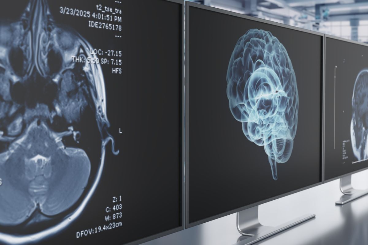
We’ve known for decades that newborn babies display a surprising fondness for faces. Even just a few hours after birth, new babies respond to voices, make eye contact, and favor faces that look back at them.
But the bigger challenge for researchers has been figuring out how the newborn brain supports that social orientation and why some infants aren’t as engaged.
A new study in Biological Psychiatry: Global Open Science unlocks a critical part of that puzzle. Researchers from Yale University and the Developing Human Connectome Project (dHCP) have discovered that a right-lateralized “social perception” pathway in the brain is already up and running at birth.
“We found that connectivity within this network was already quite robust within a couple of weeks after birth,” says senior author Dustin Scheinost, PhD, associate director of biomedical imaging technologies at the Yale Biomedical Imaging Institute.
The strength of this wiring predicts how much attention babies give to faces four months later.
Mapping the Newborn Brain
The research team relied on a pair of robust datasets for their study:
- More than 300 full-term neonates from the open-access dHCP study, scanned at roughly 41 weeks postmenstrual age; and
- And more than six dozen Yale infants, including some with a family history of autism, scanned within the first six weeks of life and followed throughout their time as toddlers.
Using resting-state functional MRI, the researchers measured intrinsic functional connectivity (iFC) across two crucial brain systems:
- The social perception pathway, which links visual areas to motion-sensitive and speech-related regions in the superior temporal sulcus (STS); and
- The ventral face pathway, which supports static face recognition in the fusiform gyrus.
In both datasets, the right and left social pathways showed healthy (and active) internal communication. But the right-sided network really stood out. It not only displayed stronger overall connectivity, but it also grew more coordinated with age, hinting that early social exposure helps fine-tune this nascent circuitry.
From Brain to Behavior
To decipher how those neural connections translate into real-world social behavior, the Yale team invited 37 infants back at four months for an eye-tracking task. The babies watched short videos of a person speaking while surrounded by distracting toys.
The results were abundantly clear. Infants born with stronger right social pathways spent a lot more time looking at faces. And that persisted even after the researchers accounted for age and motion during scanning. It also remained specific to that right-social circuit. No similar pattern emerged in the left-sided or ventral face networks.
And by 18 months, those same infants who took more time looking at faces also showed fewer social-communication concerns, as measured by parental questionnaires.
This, the researchers conclude, suggests a developmental cascade. Early brain wiring influences early attention patterns, which – in turn – form the foundation of one’s social abilities later on in life.
Autism and the Earliest Warning Signs
This revelation could lead to a big shift in autism research. Diminished attention to faces remains one of the most reliable early behavioral markers of autism spectrum disorder (ASD), usually detectable months before any kind of diagnosis.
Earlier studies have found atypical STS connectivity in older children with autism. But this is the first paper to trace the link back to the neonatal brain.
That weaker right-STS connectivity at birth, the study’s authors suggest, could signal a biological vulnerability that quietly influences how infants engage socially. Over time, that could cap social learning opportunities and add to downstream challenges in social cognition.
The study adds to a mounting body of evidence that functional brain connectivity might bridge genetics and behavior. Earlier work from the same team showed that newborns with altered insula-amygdala connectivity – another social circuit – tended to show higher autism-related traits a year later.
This new study stretches that idea to the right temporal cortex, pinpointing a circuit that might underlie early social attention itself.
Built for Connection
Even among typically developing infants, this new research highlights just how well-equipped the newborn brain is for human interaction. The right-STS network appears to be an experience-expectant system: prewired and just waiting for social input to make it better. Every coo, smile, and moment of eye contact probably helps test and strengthen that circuit.
Ultimately, the study reveals that the newborn brain is hardly a blank slate. Long before smiles, giggles, or even words, a right-sided web of cortical regions is already humming along, preparing infants to perceive – and be a part of – the social world around them.
“This work will help us understand more about the brain processes that drive social attention in typical development and that may be involved in the social vulnerabilities we know are associated with autism,” Yale’s Emily Fraser Beede Professor of Child Psychiatry and co-senior author Katarzyna Chawarska, PhD, said.
Further Reading
Two Genetic Paths Dictate Timing of Autism Diagnosis
New Autism Study Uncovers Four Biological Subtypes
Siblings With Autism Share More of Father’s DNA, Not Mother’s


