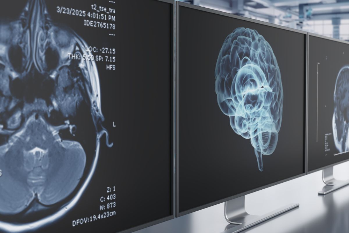
For decades, neuroscientists nurtured visions of plotting an atlas of the human brain at work. They wanted more than the collection of snapshots of isolated regions of the brain. They wanted a coordinated map charting how things like action, expectation, memory, and perception work together.
This month, a pair of companion studies out of the International Brain Laboratory (IBL) – and appearing in the journal Nature – deliver the most ambitious step yet toward that goal. Using groundbreaking new technology, including widefield calcium imaging, the researchers recorded more than 600,000 neurons across 279 brain areas in mice performing a complex decision-making task. The result? A brain-wide view of how animals integrate what they see, what they expect, and how they act.
Together, the studies reveal two startling truths:
- That neural signals for sensation, action, and reward spread far more widely than the researchers thought.
- And that the brain’s representation of “prior knowledge”— expectations built from past experience – isn’t relegated to high-level decision hubs. Instead, the authors discovered that it weaves itself all throughout the brain, from primary sensory circuits to motor regions and even the prefrontal cortex. Expectation, they argue, is baked into the system.
Mice, Wheels, and Random Odds
Both studies stemmed from the same behavioral paradigm. Researchers trained the mice to ascertain the position of a visual grating on a screen and indicate their choice by turning a small wheel with their paws.
The twist? After an initial set of unbiased trials, the probability that the stimulus appeared on the left or right shifted silently in blocks – sometimes 80:20, sometimes 20:80. To succeed, the mice had to combine immediate sensory evidence with a statistical sense of the current block’s bias.
This setup had two advantages. It was complex enough to recruit sensory, cognitive, and motor processes, yet standardized across a dozen labs around the world. That consistency meant data could be pooled, compared, and analyzed at scale. Across 139 mice, the team logged 699 Neuropixels probe insertions, generating activity traces from 621,733 neurons and covering virtually every major division of the brain.
A Census of Neural Correlates
The first paper documents the authors’ effort to catalog where in the brain different aspects of the task were encoded.
The researchers tracked four key variables:
- The visual stimulus.
- The animal’s choice.
- Motor movement (wheel turns).
- And feedback (reward or time-out).
The researchers produced predictable – yet still surprising – results. As expected, visual stimuli first sparked activity in classical visual areas, such as the primary visual cortex and thalamic relay nuclei.
But in as quickly as 100 milliseconds, signatures of the stimulus spread far and wide into midbrain and hindbrain regions. Similarly, neurons represented motor preparation and movement almost everywhere, from striatum to cerebellum. Even reward responses cropped up in dozens of different areas, stretched far beyond the dopamine-rich centers normally tied to reinforcement.
And this is what surprised the researchers the most: Just widespread these signals appeared to be. They failed to find any single, vital “decision center.” While specific regions were strongly tuned, the team found neural correlates of choice and action scattered throughout the brain. In effect, the researchers contend that decision-making is not a localized operation but a networked function – an emergent property of many areas working in parallel.
The Puzzle of Priors
If the first study mapped the “what” of decision-related signals, the second zoomed in on the “expectation” piece. In Bayesian terms, a “prior” is the brain’s estimate of the world before new evidence arrives. In this task, the prior was the mouse’s internal guess about whether the current block was left- or right-biased.
Behavioral analyses confirmed that mice relied on the block structure. On zero-contrast trials, the mice still performed well above chance, guided solely by their expectation. They also adapted gradually after block switches, taking anywhere between five and 10 trials to update their internal model. Not perfectly Bayesian, but close.
Where, then, was this prior represented neurally? By decoding neural activity during the quiet inter-trial interval, researchers could predict the Bayes-optimal prior from activity patterns in 20% to 30% of recorded brain regions. These included not only high-level association areas, such as the orbitofrontal and anterior cingulate cortex, but also early sensory regions (lateral geniculate nucleus, primary visual cortex) and deep motor structures (gigantocellular reticular nucleus, superior colliculus).
This discovery threatens a long-standing assumption that priors show up late in the decision making pipeline. Instead, expectation appears to saturate the entire brain, shaping sensory representations before stimuli ever show up.
A Loopy Network
To probe how this prior information might circulate, the team applied Granger causality analyses. They uncovered a dense, “loopy” network where prior signals flowed forward from subcortical to cortical regions, as well as back from higher-order cortex to early sensory areas.
Simply put, the system doesn’t just accumulate evidence in a straight line. It recycles expectations through recurrent loops, consistent with theoretical models of the brain as a probabilistic inference machine.
Notably, the neural prior wasn’t just a by-product of posture or eye movements. By comparing with high-speed video tracking, the researchers showed that prior decoding couldn’t be fully explained by subtle shifts in gaze or body position. The signal reflected cognition, not just embodiment.
Work Outside the Lab
Both papers stress that the real contribution isn’t the discovery that makes the headlines. It’s the dataset itself. And the IBL hosts all the recordings, behavioral data, and analysis pipelines and has made them publicly available. With single-neuron resolution across hundreds of regions, the resource invites others to test hypotheses about distributed computation, cell-type specificity, and circuit dynamics.
While the work is in mice, the broader implications are clear. The study’s authors insist that human decision-making almost certainly relies on similar principles.
“The efforts of our collaboration generated fundamental insights about the brain-wide circuits that support complex cognition; this is really exciting and a major step forward relative to the ‘piecemeal’ approach (1-2 brain areas at a time) that was previously the accepted method in the field,” IBL researchers and University of California, Los Angeles Professor of Neurobiology Dr. Anne Churchland, explained. “My hope going forward is that both our scientific discoveries and our new insights on reproducibility will have an impact in the field.”
Clinical research, too, can benefit. Disorders from schizophrenia to Parkinson’s involve disrupted integration of sensory input, expectations, and action. A better map of the healthy network could help clinicians find out how – and where – those circuits go awry.
This discovery could also offer up lessons for artificial intelligence. Many machine learning systems still treat priors as add-ons to sensory evidence. The mouse brain suggests a much more intricate design where expectations shape perception from the ground up, through recursive loops that span a massive network.
A New Cartography
Together, these two studies upend what we think we know about cognition. Decision-making emerges not from isolated cortical command centers, but from distributed, recursive interactions across sensory, motor, and association regions.
It’s an exhilarating, if humbling, revelation for neuroscientists. The brain is even messier than earlier models implied. But it’s also more elegant. It looks a lot like a Bayesian network brought to life, looping and relaying information in a way that makes perception and action inseparable. And thanks to this historic dataset, neuroscientists now have a map to help them navigate that messy terrain.
As one researcher put it, “We didn’t just map the brain during decision-making. We caught the brain in the act of thinking.”
Further Reading
Therapy Reshapes the Depressed Brain (Literally)