A new JAMA Neurology study suggests that when Alzheimer’s symptoms don’t line up the way we think they should with our documented biological markers, it can reveal who’s likely to decline quickly. It can also point to who might be more resilient than most.
But more importantly, what this international team of researchers uncovered could help clinicians make better prognoses and more quickly (and accurately) interpret treatment responses.
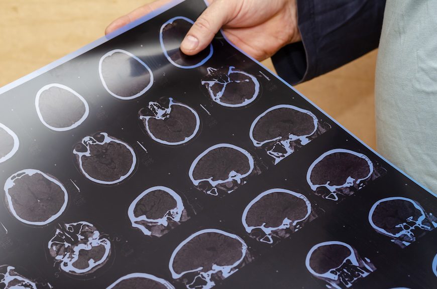
For years, clinicians have relied on biomarkers such as amyloid-β and tau to diagnose Alzheimer’s disease. But biology isn’t always forthcoming. Some patients with substantial tau pathology function much better than expected. While others suffer sharp cognitive decline despite relatively modest tau levels.
This new research focuses on that discrepancy, what researchers call “tau-clinical mismatch.”
Methodology
Led by the University of Pennsylvania’s Christopher A. Brown, MD, PhD, the research team examined data from nearly 900 amyloid-positive individuals enrolled in the Alzheimer’s Disease Neuroimaging Initiative (ADNI) and an independent Penn Alzheimer’s Disease Research Center cohort. All participants underwent detailed clinical assessments alongside measures of tau burden, obtained either through tau PET imaging or a blood-based biomarker known as phosphorylated tau 217 (p-tau217).
The researchers compared tau burden with clinical impairment, measured by the Clinical Dementia Rating Sum of Boxes (CDR-SB), and classified participants into one of three groups.
- Canonical individuals showed the expected alignment between tau and symptoms.
- Resilient individuals functioned better than their tau levels would predict.
- Vulnerable individuals showed worse cognitive impairment than expected – given their tau burden.
Those vulnerable patients stood out immediately. Across datasets, they were more likely to
show evidence of additional brain pathologies beyond classic Alzheimer’s changes. Imaging signatures pointed to greater involvement of TDP-43, a protein linked to limbic-predominant age-related TDP-43 encephalopathy. Cerebrospinal fluid tests further revealed higher rates of α-synuclein positivity, a marker associated with Lewy body disease.
Making Sense of Contradictions
In short, their symptoms appeared to be driven not just by Alzheimer’s pathology, but by a complicated mashup of neurodegenerative processes.
Structural brain imaging told a similar story. Compared with canonical patients, vulnerable individuals showed thinner medial temporal lobe and temporopolar regions. These differences persisted even after accounting for other factors, such as overall tau burden, age, sex, and education.
The mismatch also carried powerful prognostic information. When the researchers aligned participants along a biological timeline anchored to estimated tau onset, clinical trajectories diverged sharply. Vulnerable individuals declined earlier and faster, with steeper increases in CDR-SB scores and quicker drops on cognitive tests.
Resilient individuals, on the other hand, showed delayed symptom onset and slower progression. In fact, these individuals often remained somewhat for years after tau positivity.
Finally, looking closer at the survival analyses, vulnerable participants seemed to be roughly twice as likely as canonical patients to progress to the next clinical stage. Resilient individuals displayed an inverse pattern, with a much lower progression risk.
What It Might Mean
One of the study’s most practical contributions lies in its use of blood-based biomarkers. While tau PET scans remain largely confined to research settings, plasma p-tau217 is already becoming clinically available. Models based on p-tau217 performed nearly identically to those using tau PET, classifying patients into the same mismatch groups with high concordance. That opens the door to applying this framework in everyday clinical practice.
To illustrate that potential, the researchers applied their model to a real-world cohort of patients receiving anti-amyloid therapies. Even before treatment began, mismatch classification predicted markedly different expected trajectories over an 18-month period. The implication is that tau-clinical mismatch could provide individualized “null models” of decline. These benchmarks could help clinicians better judge whether a therapy is helping a particular patient.
Taken together, the findings suggest that when Alzheimer’s symptoms appear to be out of proportion to tau pathology, that imbalance isn’t just noise. It’s a signal. And in a field turning toward precision medicine, learning to read that signal could help clinicians distinguish accelerated disease from resilience, and tailor a patient’s care accordingly.
Further Reading
Benefits and Risks of New Tests for Alzheimer’s Disease
Lipid Deficit Might Explain Higher Alzheimer’s Risk In Women
Sleep Disorders Linked to Higher Risk of Dementia and Parkinson’s