aDepartment of Psychiatry and Behavioral Sciences, Eastern Virginia Medical School, Norfolk, Virginia
*Corresponding author: David R. Spiegel, MD, Department of Psychiatry and Behavior Sciences, Eastern Virginia Medical School, 825 Fairfax Ave, Norfolk, VA 23507 ([email protected]).
Prim Care Companion CNS Disord 2021;23(2):20l02742
To cite: Spiegel DR, D’Souza HS, Yee KL, et al. A case of Balint syndrome due to left basal ganglia hemorrhagic stroke: exploring the pathogenesis through parietal lobe circuits. Prim Care Companion CNS Disord. 2021;23(2):20l02742.
To share: https://doi.org/10.4088/PCC.20l02742
© Copyright 2021 Physicians Postgraduate Press, Inc.
Balint syndrome (BS) is a rare neurologic disorder associated with bilateral parieto-occipital damage that disrupts connections between the posterior visual association areas and the motor regions of the prefrontal cortex.1 It typically consists of inaccurate reach responses to objects under visual guidance (optic ataxia [OAt]), disturbed organization of eye movements (ocular apraxia [OcA]), and an inability to perceive more than one object at a time (dorsal simultanagnosia [DS]). These symptoms are thought to develop, in part, from an impairment in visual attention, although OAt may also involve a visual proprioceptive deficit.2,3 Additionally, basal ganglia (BG) participates in circuits involving visual attention vis-a-vis the fronto-parietal attention network,4 oculomotor control,5 and prehension movements.6 Thus, lesions in BG potentially could present with DS, OcA, or OAt. We present a case of BS that followed left BG hemorrhagic stroke.
Case Report
The patient was a 76-year-old woman who came to the hospital after her husband noted she was “uncoordinated.” Her vital signs were stable except for blood pressure of 223 mm Hg/152 mm Hg. Computed tomography (CT) of the brain revealed a left BG hemorrhagic infarct. The patient scored 16 on the National Institutes of Health Stroke Scale.7 Pertinent positives included right-sided gaze palsy and hemiparesis.
Her past medical history was significant for hypertension, although she was not taking medications prior to admission. She had a remote history of nicotine and alcohol usage. Blood alcohol level and urine drug screen were negative. Her husband denied previous history of motor or cognitive deficits. Due to “restlessness,” magnetic resonance imaging (MRI) was unobtainable until hospital day 30, but it corroborated CT findings and revealed additional hematoma encompassing left dorsal BG.
After the patient was reported removing her nasogastric tube, we were consulted on hospital day 35. Mental status examination was remarkable for dysarthria, although our patient was able to follow instructions without difficulty. When asked to track an object with her eyes, the patient had smooth saccadic movements to the left, but kept turning her head while trying to track objects to the right visual field. Her Mini-Mental State Examination8 score was 21, but she had a negative assessment per the Confusion Assessment Method.9 Given improvement in right hemiparesis, we evaluated our patient for BS.
When presented with the “cookie theft” picture,10 our patient was able to identify individual items but could not describe the scene as a whole. She was shown a Navon figure (characters composed of smaller characters),11 wherein she fixated on smaller letters and failed to report the larger letter E when prompted. Our patient was unable demonstrate voluntary right visual field saccades (left-sided saccades were unaffected). Nonetheless, 2 weeks later, difficulty producing voluntary saccades to verbal commands resolved. Finally, per the protocol of Borchers and colleagues,12 misreaching behavior (she neither grasped the target in the first nor had other following movement) was observed using her contralesional arm for movements directed toward the contralesional/right visual half-field.
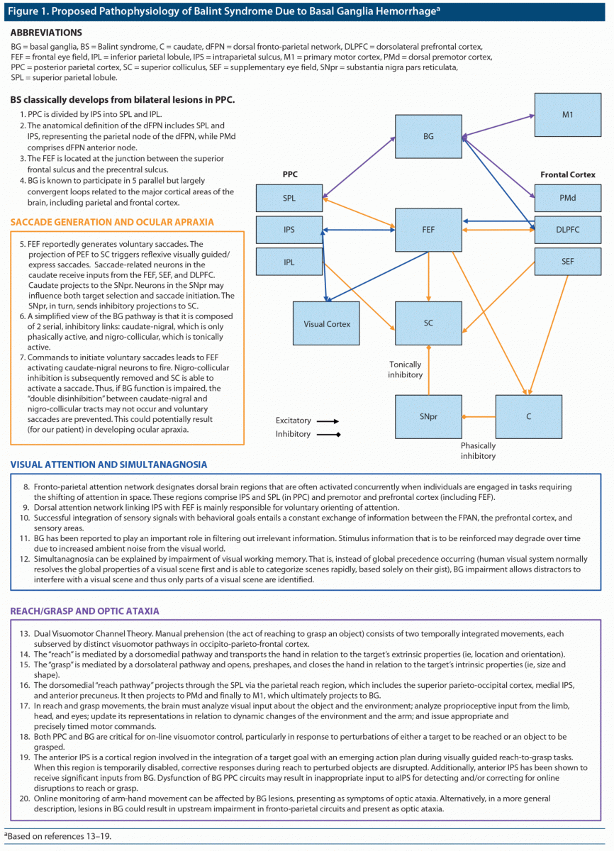
On the basis of this evaluation, we believed our patient’s phenotype included DS, OcA, and OAt. For a review of BS in our patient, please see Figure 1.13–19
Discussion
In conclusion, similar to other neuropsychiatric disease, our patient’s case of BS due to BG lesion can plausibly be viewed as a “circuit disorder.”20 BG is structurally represented within and between fronto-parietal attention (posterior parietal cortex [PPC]/dorsal frontal cortex including frontal eye fields), oculomotor (frontal/parietal oculomotor cortex), and dorsomedial reach pathway (primary motor cortex/supplementary motor area and PPC/superior parietal lobule) circuits.16,18,21–23 While BS has overwhelmingly been associated with parieto-occipital pathology, different lesion combinations have been described in the literature.24,25 Thus, per our case, a BG lesion within or between these 3 parietal lobe circuits could present with the phenotype of BS, ie, DS, OcA, and OAt, respectively, yet without the classical bilateral parieto-occipital lesions that typically produce BS.
Published online: April 1, 2021.
Potential conflicts of interest: Dr Spiegel is in the Speaker’s Bureau for Allergen, Alkermers, Otsuka, and IntraCellular Pharmaceuticals, but has no conflict of interest in preparation of this manuscript. Drs D’Souza, Yee, Kot, Yuna, Samaras, and Cook have no disclaimer/conflict of interest to report.
Funding/support: None.
Patient consent: The patient (and husband) did give verbal consent to publish the manuscript, and we have de-identified her to protect anonymity.
References (25)

- Drane DL, Lee GP, Huthwaite JS, et al. Development of a partial Balint’s syndrome in a congenitally deaf patient presenting as pseudo-aphasia. Clin Neuropsychol. 2009;23(4):715–728. PubMed CrossRef NLM
- Zgaljardic DJ, Yancy S, Levinson J, et al. Balint’s syndrome and post-acute brain injury rehabilitation: a case report. Brain Inj. 2011;25(9):909–917. PubMed CrossRef NLM
- Jacobs DA, Liu GT, Nelson PT, et al. Primary central nervous system angiitis, amyloid angiopathy, and Alzheimer’s pathology presenting with Balint’s syndrome. Surv Ophthalmol. 2004;49(4):454–459. PubMed CrossRef NLM
- Henry MJ, Herrmann B, Obleser J. Selective attention to temporal features on nested time scales. Cereb Cortex. 2015;25(2):450–459. PubMed CrossRef NLM
- Lanciego JL, Luquin N, Obeso JA. Functional neuroanatomy of the basal ganglia. Cold Spring Harb Perspect Med. 2012;2(12):a009621. PubMed CrossRef NLM
- Grafton ST. The cognitive neuroscience of prehension: recent developments. Exp Brain Res. 2010;204(4):475–491. PubMed CrossRef NLM
- NIH Stroke Scale. National Institute of Neurological Disorders and Stroke. October 1, 2003. https://www.ninds.nih.gov/sites/default/files/nih_stroke_scale_508c.pdf
- Folstein MF, Folstein SE, McHugh PR. “Mini-mental state”: a practical method for grading the cognitive state of patients for the clinician. J Psychiatr Res. 1975;12(3):189–198. PubMed CrossRef NLM
- Inouye SK, van Dyck CH, Alessi CA, et al. Clarifying confusion: the confusion assessment method: a new method for detection of delirium. Ann Intern Med. 1990;113(12):941–948. PubMed CrossRef NLM
- Goodglass H, Kaplan E. The Assessment of Aphasia and Related Disorders. Philadelphia, PA: Lea & Febiger; 1983.
- Navon D. Forest before trees: the precedence of global features in visual perception. Cognit Psychol. 1977;9(3):353–383. CrossRef
- Borchers S, Müller L, Synofzik M, et al. Guidelines and quality measures for the diagnosis of optic ataxia. Front Hum Neurosci. 2013;7:324. PubMed CrossRef NLM
- Bisley JW, Goldberg ME. Attention, intention, and priority in the parietal lobe. Annu Rev Neurosci. 2010;33(1):1–21. PubMed CrossRef NLM
- Ptak R, Schnider A, Fellrath J. The dorsal frontoparietal network: a core system for emulated action. Trends Cogn Sci. 2017;21(8):589–599. PubMed CrossRef NLM
- Su CY, Chen HM, Kwan AL, et al. Neuropsychological impairment after hemorrhagic stroke in basal ganglia. Arch Clin Neuropsychol. 2007;22(4):465–474. PubMed CrossRef NLM
- Leigh RJ, Kennard C. Using saccades as a research tool in the clinical neurosciences. Brain. 2004;127(pt 3):460–477. PubMed CrossRef PubMed CrossRef NLM
- Ptak R. The frontoparietal attention network of the human brain: action, saliency, and a priority map of the environment. Neuroscientist. 2012;18(5):502–515. PubMed CrossRef NLM
- Voytek B, Knight RT. Prefrontal cortex and basal ganglia contributions to visual working memory. Proc Natl Acad Sci U S A. 2010;107(42):18167–18172. PubMed CrossRef NLM
- Thomas C, Kveraga K, Huberle E, et al. Enabling global processing in simultanagnosia by psychophysical biasing of visual pathways. Brain. 2012;135(Pt 5):1578–1585. PubMed CrossRef NLM
- DeLong M, Wichmann T. Changing views of basal ganglia circuits and circuit disorders. Clin EEG Neurosci. 2010;41(2):61–67. PubMed CrossRef NLM
- Karl JM, Whishaw IQ. Different evolutionary origins for the reach and the grasp: an explanation for dual visuomotor channels in primate parietofrontal cortex. Front Neurol. 2013;4:208. PubMed CrossRef NLM
- Inouchi M, Matsumoto R, Taki J, et al. Role of posterior parietal cortex in reaching movements in humans: clinical implication for ‘optic ataxia.’ Clin Neurophysiol. 2013;124(11):2230–2241. PubMed CrossRef NLM
- Lukos JR, Snider J, Hernandez ME, et al. Parkinson’s disease patients show impaired corrective grasp control and eye-hand coupling when reaching to grasp virtual objects. Neuroscience. 2013;254:205–221. PubMed CrossRef NLM
- Chechlacz M, Humphreys GW. The enigma of Bálint’s syndrome: neural substrates and cognitive deficits. Front Hum Neurosci. 2014;8:123. PubMed CrossRef NLM
- Rizzo M, Vecera SP. Psychoanatomical substrates of Bálint’s syndrome. J Neurol Neurosurg Psychiatry. 2002;72(2):162–178. PubMed CrossRef NLM
Enjoy this premium PDF as part of your membership benefits!