Because this piece does not have an abstract, we have provided for your benefit the first 3 sentences of the full text.
Have you ever wondered what is responsible for an altered mental status with abnormal movements? Have you been uncertain about when and how to evaluate a patient with such signs and symptoms? Have you been perplexed by how to treat such an individual over the short and long term?
From the Editors

Are you a healthcare provider?
Add your NPI to personalize your JCP experience.
Catatonia:
An Approach to Diagnosis and Treatment
Have you ever wondered what is responsible for an altered mental status with abnormal movements? Have you been uncertain about when and how to evaluate a patient with such signs and symptoms? Have you been perplexed by how to treat such an individual over the short and long term? If you have, then the following case vignette and discussion (which illustrates the complexity of diagnosing and treating a patient with a mixture of motor and psychological symptoms) should prove useful.
CASE VIGNETTE: PART 1
Ms A, a 53-year-old woman, presented to the emergency department of a community hospital with altered mental status, weakness, and unusual movements. She had been calling emergency medical services frequently for 5 days prior to her admission complaining of recurrent falls. Her medical history was notable for type 2 diabetes mellitus, osteoarthritis (status post right knee fusion), asthma, anemia, recurrent urinary tract infections, and a Roux-en-Y gastric bypass. Her psychiatric history included major depressive disorder, posttraumatic stress disorder, borderline personality disorder, opiate use disorder, and a recent diagnosis of conversion disorder (manifested by stuttering speech and shaking movements). Significant laboratory abnormalities included elevated liver enzymes, an elevated creatine kinase level, severe hypokalemia, and an electrocardiogram that showed a prolonged QTc (650 ms; reference range, ≤ 450 ms for women). She was subsequently transferred to our hospital for additional workup.
At admission, Ms A was disoriented and somnolent. Collateral information revealed several weeks of poor oral intake and recent psychosocial stressors, including her son’s graduation from high school and the pending transfer of care from her therapist of nearly 4 years to a new provider. Her developmental history included physical and sexual abuse in childhood, and her psychiatric history involved multiple psychiatric hospitalizations, prior suicide attempts, and self-injurious behaviors (including head banging and wrist cutting). Ms A’s substance use history was significant for opioid use disorder actively managed with medication-assisted treatment (buprenorphine/naloxone). Her family history was positive for alcohol use disorder, and her father had committed suicide. Her medication list included cyproheptadine, doxepin, lamotrigine, eszopiclone, hydroxyzine, sertraline, melatonin, buprenorphine/naloxone, insulin glargine, fluconazole, omeprazole, ranitidine, polyethylene glycol, and meloxicam.
Ms A was afebrile with a blood pressure of 147/77
mm Hg and a heart rate of 94 bpm. Her respiratory function was normal. Laboratory tests included hemoglobin: 11.1 g/dL (reference range, 12.0–15.2 g/dL), mean corpuscular volume: 75.6 fL (reference range, 82–99 fL), potassium: 2.1 mEq/L (reference range, 3.5–5.0 mEq/L), albumin: 1.9 g/dL (reference range, 3.2–5.0 g/dL), pre-albumin: 17 mg/dL (reference range, 18–38 mg/dL), aspartate aminotransferase: 137 U/L (reference range, 7–52 U/L), alanine aminotransferase: 88 U/L (reference range, 5–34 U/L), and creatine kinase: 1,531 U/L (reference range, 30–200 U/L). Her ammonia and B12 levels were within normal limits. An infectious and toxicology workup was negative. Her folate level was low. Magnetic resonance imaging (MRI) scan of the brain was within normal limits. An electroencephalogram (EEG) was not performed, as there was a low level of clinical suspicion for a seizure disorder.
The examination revealed no flushing or diaphoresis. She was intermittently somnolent and agitated, with prominent non–goal-directed motor activity (ie, shoulder and leg-shaking movements). Her eye contact was poor, with bizarre eye-rolling movements. Her speech was sparse, incoherent, and slurred. She reported a “depressed” mood and had a blunted affect. She endorsed suicidal ideation with no plan and suspiciousness. Her thought process was disorganized, with loosening of associations, word salad, and thought blocking. She reported having visual hallucinations of snakes. She was oriented to self, date, day, and year, but not to place despite multiple attempts at correction.
DISCUSSION
What Is Catatonia?
Catatonia is a fascinating clinical phenomenon that occurs not only in patients with psychiatric illness (eg, mood disorders, schizophrenia) but also in those with neurologic diseases and other medical conditions.1 Signs and symptoms of catatonia include excitement, immobility/stupor, mutism, staring, grimacing, stereotypy, mannerisms, rigidity, negativism, and withdrawal.2 Patients with the catatonic syndrome may also exhibit posturing/catalepsy, echopraxia/echolalia (ie, mimicking of examiner’s movements/speech), verbigeration (ie, repetition of phrases or sentences like a scratched record), waxy flexibility, impulsivity, automatic obedience, mitgehen (“Anglepoise lamp” arm raising in response to light pressure of finger, despite instruction to the contrary), gegenhalten (resistance to passive movement, which is proportional to strength of stimulus and appears automatic rather than willful), ambitendency (when the patient appears motorically “stuck” in indecisive, hesitant movement), grasp reflex, perseveration, combativeness, and autonomic abnormality.
Signs and symptoms of catatonia most likely reflect underlying neurobiology. Indeed, catatonia represents an overlapping neurocircuitry that is complicated but has discernable connections (G. Fricchione, MD, personal communication, September 21, 2017). Catatonia is a potentially life-threatening syndrome. In stuporous catatonia, refusal to eat or drink may lead to dehydration and cardiovascular collapse. In hyperactive catatonia, excessive motor activity may lead to hyperthermia, seizures, or death.
How Is Catatonia Diagnosed?
The Bush-Francis Catatonia Rating Scale2 has been helpful as a clinical tool to identify catatonia and to differentiate catatonia from other neuropsychiatric phenomena. Criteria for catatonia were simplified during the transition from the DSM-IV3 to the DSM-54 to enhance the recognition of catatonia and encourage its specific treatment. Situations may arise in which the underlying cause of catatonia (eg, brief psychotic episode, schizophrenic disorder, schizoaffective disorder, substance-induced psychotic disorder) for a patient is not immediately recognized, and the DSM-5 category of catatonia, not otherwise specified, now allows for the diagnosis to be made.1
Which Conditions Share
Common Features With Catatonia?
Patients who are exhibiting catatonic signs (ie, appear to be “playing possum”) can be categorized into 2 distinct groups: those who cannot interact with the examiner due to avolitional causes and those who will not interact with the examiner by voluntary choice.5 Those who cannot participate in a clinical interview may have conversion disorder (ie, unusual or impaired motor or sensory function with no medical or neurologic cause, and the symptoms are not feigned by the patient), akinetic mutism, coma, locked-in syndrome, delirium, seizure, or a dissociative state. Patients who will not interact include individuals who are in pursuit of tangible secondary gain (eg, shelter, avoidance of legal or social obligations) or those with factitious disorders wherein the primary gain is to assume the sick role and obtain medical care. Patients who meet criteria for the diagnosis of catatonia would fit into the “cannot interact” category.
How Has Our Understanding
of Catatonia Evolved Over Time?
Kahlbaum6 (1828–1899) first conceptualized catatonia as a neurologic condition with psychiatric manifestations, while recognizing its protean forms and etiologies. Expressions of the disease (or symptom-complexes) included melancholia (eg, withdrawal, psychomotor retardation, and mutism), mania (eg, hyperkinetic, exalted, and verbigerating), atonia (eg, posturing, convulsions, and flexibilitas cerea), and dementia (eg, terminal stage in which the patient descends into apathy and mental poverty).6–8 Kahlbaum’s disciple, Hecker, in 1871 wrote that catatonia (or melancholia attonita) had a “characteristic course including a series of nosologically important symptoms leading to a clear delineation as a disease entity.”9(p358)
Unfortunately, Kraepelin (1859–1926), who like Kahlbaum was a pioneer of descriptive psychiatry, subsumed catatonia under the category of dementia praecox, which was later renamed schizophrenia by Bleuler (1857–1939).10 This misguided “catatonia = schizophrenia” equation has persisted in modern medicine and put patients at risk for being administered dopamine antagonists (used to treat schizophrenia); these agents have precipitated neuroleptic malignant syndrome in patients with catatonia. Signs and symptoms of catatonia are frequently overlooked in those who do not suffer from schizophrenia. Moreover, specific and effective treatments for catatonia, which are different from standard treatments of schizophrenia, are neglected.
Another misstep in our understanding of catatonia as a syndrome occurred when Bleuler, who emphasized the psychological underpinnings of catatonia at the expense of neuromedical explanations, viewed patients’ abnormal postures, rigidity, and stupor as purely psychological defense mechanisms designed to “shut out reality.”11–13
Gelenberg14 attempted to untether catatonia from its moorings as a subtype of schizophrenia with purely psychological origins and reiterated Kahlbaum’s conceptualization of catatonia as a syndrome with multiple manifestations. Gelenberg and Mandel15 elegantly described catatonia as a biological response to dopamine blockade that may represent a psychosocial withdrawal symptom to an altered motor system.
Indeed, the rigidity often present in catatonia (primarily considered a biological/motor response to dopamine blockade) renders animals unable to move. The related behavior of playing possum,5 a psychological response often observed in traumatized or otherwise threatened individuals, has been suggested to play an adaptive role in maintaining survival and highlights catatonia’s postulated evolutionary roots and its position at the interface of biology and psychology.
What Causes Catatonia?
The nervous system evolved in order to mobilize (via the sympathetic nervous system) or immobilize animals (via the parasympathetic nervous system).16 Although catatonic immobilization or “freezing” in the face of danger may seem counterintuitive, tonic immobility may be the best option when there seems to be little immediate chance of escape or of winning a fight. Indeed, the so-called “death feign” among animals has served as a passive defense against predators by which potential prey can evade the visual display and motion-tracking of predators.17
Jackson18 postulated that recently developed evolutionary innovations are attempted first in times of stress. In affective and thought disorders, the more evolved ventral vagal complex, or smart vagus,19 may trigger disinhibited sympathetic mobilization. If this mechanism fails, the next step would be a more primitive “vegetative” vagal immobilization response mediated by the dorsal vagal complex or playing possum in a traditional catatonic withdrawal state.
Catatonia may be viewed as a disorder of the basal ganglia–thalamocortical circuits of Alexander and DeLong.20,21 The thalamus, as the sensory node, senses the external environment and sends afferent fibers to the amygdala (via a rapid one-synapse passage) and to nodes in cortical terminal zones, which in turn send corticostriatal afferents for salience to the ventral striatum and for motor processing to the dorsal striatum, which then enact motor programs and also recontact the thalamus to control the thalamic filtering of information destined to reach cortical areas in thalamocortical tracts. These circuits serve to sense the environment, analyze incoming data, create internal brain representations of the external state, and construct a premotor template of responses to effect mobilization/immobilization (ie, approach/avoidance).16
Lesions of the dorsolateral prefrontal cortex or, importantly, anywhere along the basal ganglia–thalamocortical loop known as the dorsolateral prefrontal circuit cause executive dysfunction.22 Therefore, diffuse periventricular white matter changes on brain MRI could represent disruption in important basal ganglia–thalamocortical circuits and may predispose an individual to catatonia.
Why Does Catatonia Present With Motor Signs?
Patients with catatonia exhibit posturing wherein they show a specific, uncomfortable, and often bizarre position of parts of their body against gravity.23 They may remain with complete akinesia for hours, days, or weeks (or even years). Posturing is defined as a position taken actively and internally by the patient, while catalepsy involves a position induced passively and externally by the examiner. Posturing may occur in eyes (staring), head (psychic pillow), and limbs (classic posturing).23 Neuroimaging studies implicate right parietal dysfunction in both catatonia24 and Cotard’s syndrome,25 a nihilistic delusion that one is dead (or, paradoxically immortal), has lost his/her soul, is without functional body systems, is rotting internally, or is without limbs.26,27
Adding further to the complexity of the pathogenesis of catatonia, disturbances in important neurotransmitters in key brain areas (including the limbic or anterior-cingulate medial orbitofrontal circuit) appear to play a role.16 Specifically, imbalance of dopamine and γ-aminobutyric acid (GABA) in medial forebrain bundle dopamine tracts may set off a catatonic response.28 The medial forebrain bundle is an important neural pathway with axons running from monoaminergic nuclei in the upper brainstem to both paralimbic and frontal cortical regions. These mesolimbic/mesocortical tracts distribute the neurotransmitters serotonin (originating from the nucleus raphe), dopamine (from the ventral tegmentum), and norepinephrine (from the locus coeruleus) to the important terminal zones along the brain reward/brain motivation superhighway.29 The dopamine hypothesis of psychosis postulates that psychotic symptoms are related to hyperdopaminergia in the mesolimbic dopamine pathway (ie, dopamine tracts from the ventral tegmental area of the midbrain to the limbic system).30 Friedhoff’s31 restitutive hypothesis states that, during psychotic states, the mesolimbic system will adjust itself to attempt restoration of homeostasis. The brain, in essence, compensates for the hyperdopaminergic state of psychosis by down-regulating dopamine receptors with the goal of reaching equilibrium. Unfortunately, this down-regulation of dopamine receptors does not limit itself to the mesolimbic system. These effects also occur in other brain systems (eg, hypothalamus, mesostriatum) in which the down-regulation can be potentially harmful. If these areas are hypodopaminergic to a large degree, yet psychosis continues, then catatonia may emerge. Moreover, this compensatory down-regulation of dopamine receptors increases vulnerability to development of catatonia with the addition of dopamine-blocking agents designed to lessen psychotic symptoms.31 Conceptualizations of catatonia as nothing more than a variant of schizophrenia are indeed dangerous for patients, as dopamine-blocking agents compound, rather than improve, the problem of the catatonic hypodopaminergic state.
What Is the Differential Diagnosis of Catatonia?
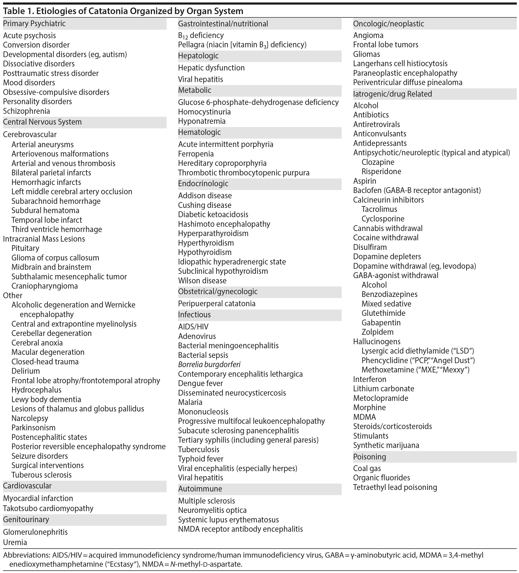
Clinicians must be vigilant for neuromedical etiologies of catatonia, as it is a syndrome found among patients with infections, endocrine disorders, metabolic derangements, and neurologic disorders.32 Thus, proper diagnosis and treatment of catatonia, and altered mental status on the whole, requires the full utilization of the physician’s medical and mental health training background.
Table 1 includes a list of conditions28,33 associated with the catatonic syndrome and is organized systematically by organ system to show the wide differential diagnosis of catatonia. These diverse, and frequently overlapping, etiologies presumably lead to catatonia vis à vis disruption of the crucial integrated, yet segregated, basal ganglia– thalamocortical loops (“the wires”) and neurotransmitters essential to the communications between nodes of these circuits (“the juices”).
What Is the Workup For Catatonia?
A neuromedical workup is critical to make clear potential etiologies of catatonia, particularly when the patient’s history corresponds with those conditions associated with catatonia (see Table 1).28 Neuroimaging (ie, MRI) should be obtained, although a negative result does not rule out neuromedical etiologies as there are toxic, metabolic, and infectious etiologies that require further scrutiny. EEG studies can be of use to diagnose epilepsy as a cause of catatonic symptoms. Lumbar puncture should be considered, along with an assay for autoantibodies (eg, those directed against the N-methyl-d-aspartate [NMDA] receptor or voltage-gated K+ channels). In malignant catatonia and neuroleptic malignant syndrome, creatine phosphokinase levels are often elevated, while serum iron levels are often reduced, and leukocytosis is frequently present (G. Fricchione, MD, written communication, July 1, 2017). In simple catatonia, serum iron reductions, creatine phosphokinase elevations, and leukocytosis can also be present.
How Can Catatonia Be Managed?
Early recognition is important in catatonia, as are close observation and frequent measurement of vital signs.34 Supportive care for patients with catatonia consists of hydration, nutrition, mobilization, anticoagulation (to prevent thrombophlebitis), and aspiration precautions. Neuroleptics and other dopamine depleters should be discontinued, and recently withdrawn dopamine agonists may need to be restarted. If hyperthermia or autonomic instability emerge or if malignant catatonia is suspected, intensive care unit supportive measures should be initiated. Clinicians should maintain a high index of suspicion for development of medical complications and new medical problems. As soon as catatonia is diagnosed, obtaining consent for electroconvulsive therapy (ECT) is a prudent measure given the possibility of progression to malignant catatonia, which is associated with significant morbidity and mortality if ECT is not administered expeditiously.34
Neurochemical studies in catatonia have concentrated primarily on the neurotransmitters GABA and dopamine, as there are clinical similarities between catatonia and basal ganglia disorders in which they are implicated.35 Lorazepam (with its muscle relaxant properties) and amantadine may relieve catatonia by changing the motor symptoms, thereby eliminating a stimulus for the withdrawal behavior.36 Benzodiazepines also lead to disinhibition. In catatonia, GABA-A agonists (eg, zolpidem) could be restorative by inhibiting sensitive pars reticulata inhibitory GABA-B neurons, resulting in disinhibition of neighboring pars compacta dopamine cells and resultant striatal dopamine agonism.28 Benzodiazepine receptors linked to GABA-A sites disinhibit mesostriatal dopamine tracts, leading to reopening (ie, widening) of the thalamic filter37 and lysing of the catatonia. Benzodiazepines may cause lysis of catatonia through effects not only on GABA receptors in the substantia nigra but also in the prefrontal cortex where corticostriatal glutamate fibers may be inhibited.28
Intravenous (IV) lorazepam is the first-line treatment for catatonia, as it is easy to administer and often helpful in diagnosis and treatment of the catatonic syndrome. An IV lorazepam challenge typically starts with 2 mg of IV lorazepam and observation for effect (G. Fricchione, MD, written communication, July 1, 2017). If there is no effect, the same dose may be repeated in 3 hours, and it can be done again in 3 hours if necessary. At least 6 mg should be given over the course of 24 hours. For those patients who show partial improvement on the first day of treatment, our recommendation is to continue 2 mg of IV lorazepam every 8 hours for at least 3 to 4 days, and it may be continued longer to maintain improvement, particularly if catatonia has been present more than a month. After full lysis of the catatonia, the patient can be switched to a sublingual or oral formulation, which typically will maintain the treatment effect. The risk/benefit profile should always be considered, and monitoring for respiratory depression is essential with benzodiazepine medication treatment.
The effectiveness of ECT may relate to its direct and indirect actions on GABA and dopamine.35,36 ECT may be given daily if necessary (ie, in more severe cases) and then at a more customary frequency (ie, every other day) once the catatonic syndrome begins to abate. Many clinicians prefer bitemporal ECT for catatonia to ensure effectiveness (G. Fricchione, MD, written communication, July 1, 2017). Maintenance ECT may be necessary in some cases.
CASE VIGNETTE: PART 2
We contemplated mixed delirium (ie, featuring components of hypoactive and hyperactive delirium), Wernicke’s encephalopathy, and seizure in our differential diagnosis. Ms A had exhibited variable speech difficulty on a prior inpatient hospitalization and was diagnosed with probable conversion disorder, and this diagnosis was also considered. We thought about the diagnosis of factitious disorder (ie, playing the “sick role”), as the patient may have pursued medical care and hospitalization to help cope with her abandonment issues surrounding the termination of treatment with her therapist and her son’s increasing independence. However, given her behavioral withdrawal (ie, decreased oral intake and sociability) and purposeless psychomotor excitement, it was our clinical judgment that catatonia was the most probable diagnosis. Ms A’s total score of 15 on the Bush-Francis Catatonia Rating Scale, with 12 elements present out of a total of 23, was a positive screening test for catatonia, which supported our diagnosis.
Ms A was administered IV lorazepam (1 mg) with the goal of lysing the catatonia. We elected to start at a lower-than-usual dose for the lorazepam challenge given the patient’s intermittent drowsiness, thin body habitus, and underlying neurodegenerative changes (ie, mild generalized volume loss on CT brain scan) that predisposed her to the development of medication side effects. Initially, she became even more somnolent, sleeping soon after receiving the lorazepam at noon until the following morning. She was remarkably more interactive during the follow-up examination (eg, she was awake, alert, and talking on the phone). Her eye contact was significantly improved, and she appeared attentive, engaged, and cooperative throughout the interview. Her speech was normal, without the stuttering or hesitation displayed earlier. She was oriented to person, place, and situation but remained disoriented to date. Her marked response to the lorazepam challenge confirmed our diagnosis of catatonia. The following day, her thiamine level was noted to be < 7 (reference range, 8–30), and it was repleted. Clinical improvement persisted into the following week of hospitalization. She was eventually discharged home with close medical and psychiatric follow-up.
SUMMARY
We described the case of a woman who developed catatonia in the context of significant interpersonal stressors as well as multiple acute and chronic medical problems to illustrate the biopsychosocial complexity from which catatonia typically arises. As demonstrated by our illustrative case, psychodynamic and psychosocial etiologies (eg, history of childhood abuse) can clearly play a contributory role in the pathogenesis of catatonic states.38 We reviewed the broad differential diagnosis of the catatonic syndrome and described treatments that have been shown to lyse catatonic states and relieve patient suffering, presumably by reregulating neurochemical imbalances.
Submitted: July 26, 2017; accepted September 28, 2017.
Published online: January 4, 2018.
Potential conflicts of interest: Dr. Stern is an employee of the Academy of Psychosomatic Medicine and has received royalties from Elsevier and the Massachusetts General Hospital Psychiatry Academy. Drs Rustad, Landsman, Ivkovic, and Finn report no conflicts of interest related to the subject of this article.
Funding/support: None.
Previous presentation: The material in this article is based on a Geisel School of Medicine at Dartmouth Psychiatry Grand Rounds presentation entitled “Catatonia in the General Hospital,” given by Drs Rustad and Landsman at Dartmouth-Hitchcock Medical Center on October 25, 2016, in Lebanon, New Hampshire.
Acknowledgment: The authors thank Gregory L. Fricchione, MD (Massachusetts General Hospital and Harvard Medical School, Boston, Massachusetts), for his helpful edits and critique of the manuscript. Dr Fricchione reports no conflicts of interest related to the subject of this article.
REFERENCES
1. Tandon R, Heckers S, Bustillo J, et al. Catatonia in DSM-5. Schizophr Res. 2013;150(1):26–30. PubMed CrossRef
2. Bush G, Fink M, Petrides G, et al. Rating scale and standardized examination. Acta Psychiatr Scand. 1996;93(2):129–136. PubMed CrossRef
3. American Psychiatric Association. Diagnostic and Statistical Manual for Mental Disorders. Fourth Edition. Washington, DC: American Psychiatric Association; 1994.
4. American Psychiatric Association. Diagnostic and Statistical Manual for Mental Disorders. Fifth Edition. Washington, DC: American Psychiatric Association; 2013.
5. Beach SR, Stern TA. “Playing possum:” differential diagnosis, workup, and treatment of profound interpersonal withdrawal. Psychosomatics. 2011;52(6):560–562. PubMed CrossRef
6. Kahlbaum K. Catatonia. Levij Y, Priden T (trans.). Baltimore, MD: Johns Hopkins University Press; 1973.
7. Quinn DK. Kahlbaum’s forms of catatonia. Psychosomatics. 2013;54(5):504–505. PubMed CrossRef
8. Azzam PN, Gopalan P. Prototypes of catatonia: diagnostic and therapeutic challenges in the general hospital. Psychosomatics. 2013;54(1):88–93. PubMed CrossRef
9. Kraam A. On the origin of the clinical standpoint in psychiatry, Dr Ewald Hecker in Görlitz. Hist Psychiatry. 2004;15(59 pt 3):345–360. PubMed
10. Fink M. Rediscovering catatonia: the biography of a treatable syndrome. Acta Psychiatr Scand suppl. 2013;127(441):1–47. PubMed CrossRef
11. Rogers DR. Catatonia: a contemporary approach. J Neuropsychiatry Clin Neurosci. 1991;3(3):334–340. PubMed CrossRef
12. Bleuler E. Dementia Praecox or the Group of Schizophrenias. Zinkin J (trans.). New York, NY: International Universities Press; 1950.
13. Bleuler E. The physiogenic and psychogenic in schizophrenia. Am J Psychiatry. 1930;110(2):203–211. CrossRef
14. Gelenberg AJ. The catatonic syndrome. Lancet. 1976;1(7973):1339–1341. PubMed CrossRef
15. Gelenberg AJ, Mandel MR. Catatonic reactions to high-potency neuroleptic drugs. Arch Gen Psychiatry. 1977;34(8):947–950. PubMed CrossRef
16. Fricchione GL. Brain evolution and the meaning of catatonia. In: Caroff SN, Mann SC, Francis A, et al, eds. Catatonia: From Psychopathology to Neurobiology. Washington, DC: American Psychiatric Publishing, Inc; 2004:201–221.
17. Schmidt NB, Richey JA, Zvolensky MJ, et al. Exploring human freeze responses to a threat stressor. J Behav Ther Exp Psychiatry. 2008;39(3):292–304. PubMed CrossRef
18. Jackson JH. Evolution and dissolution of the nervous system. In: Taylor J, ed. Selected Writings of John Hughlings Jackson. London, UK: Stapes; 1958:45–118.
19. Porges SW. Love: an emergent property of the mammalian autonomic nervous system. Psychoneuroendocrinology. 1998;23:837–861. PubMed CrossRef
20. Alexander GE, DeLong MR, Strick PL. Parallel organization of functionally segregated circuits linking basal ganglia and cortex. Annu Rev Neurosci. 1986;9(1):357–381. PubMed CrossRef
21. Alexander GE, Crutcher MD, DeLong MR. Basal ganglia-thalamocortical circuits: parallel substrates for motor, oculomotor, “prefrontal” and “limbic” functions. Prog Brain Res. 1990;85:119–146. PubMed CrossRef
22. Cummings JL. Frontal-subcortical circuits and human behavior. Arch Neurol. 1993;50(8):873–880. PubMed CrossRef
23. Mann SC, Caroff SN, Fricchione G, et al. Central dopamine hypoactivity and the pathogenesis of neuroleptic malignant syndrome. Psychiatr Ann. 2000;30(5):363–374. CrossRef
24. Northoff G, Steinke R, Nagel D, et al. Right lower prefronto-parietal cortical dysfunction in akinetic catatonia: a combined study of neuropsychology and regional cerebral blood flow. Psychol Med. 2000;30(3):583–596. PubMed CrossRef
25. Simpson P, Kaul E, Quinn D. Cotard’s syndrome with catatonia: a case presentation and discussion. Psychosomatics. 2013;54(2):196–199. PubMed CrossRef
26. Pearn J, Gardner-Thorpe C. Jules Cotard (1840–1889): his life and the unique syndrome which bears his name. Neurology. 2002;58(9):1400–1403. PubMed CrossRef
27. Ramirez-Bermudez J, Aguilar-Venegas L, Crail-Melendez D, et al. Cotard’s syndrome in neurological and psychiatric patients. J Neuropsychiatry Clin Neurosci. 2010;22(4):409–416. PubMed CrossRef
28. Fricchione G, Bush G, Fozdar M, et al. Recognition and treatment of the catatonic syndrome. J Intensive Care Med. 1997;12(3):135–147. CrossRef
29. Fricchione GL. Catatonia: a disorder of motivation and movement. Behav Brain Sci. 2002;25(5):584–585. CrossRef
30. Stahl SM. Psychosis and Schizophrenia. In: Stahl SM, ed. Essential Psychopharmacology: Neuroscientific Basis and Practical Applications. 4th ed. Cambridge, UK: Cambridge University Press; 2013:79–128.
31. Friedhoff AJ. A strategy for developing novel drugs for the treatment of schizophrenia. Schizophr Bull. 1983;9(4):555–562. PubMed CrossRef
32. Fink M, Fricchione G, Rummans T, et al. Catatonia is a systemic medical syndrome. Acta Psychiatr Scand. 2016;133(3):250–251. PubMed CrossRef
33. Denysenko L, Freudenreich O, Philibrick K, et al. Catatonia in Medically Ill Patients: An Evidence-Based Medicine (EBM) Monograph for Psychosomatic Medicine Practice. http://www.eapm.eu.com/tl_files/content/Publications/Catatonia_APM-EAPM_2015-04-17.pdf. Accessed December 12, 2017.
34. Philbrick KL, Rummans TA. Malignant catatonia. J Neuropsychiatry Clin Neurosci. 1994;6(1):1–13. PubMed CrossRef
35. Francis A. Catatonia: diagnosis, classification, and treatment. Curr Psychiatry Rep. 2010;12(3):180–185. PubMed CrossRef
36. Fricchione GL. Neuroleptic catatonia and its relationship to psychogenic catatonia. Biol Psychiatry. 1985;20(3):304–313. PubMed
37. Carlsson M, Carlsson A. Interactions between glutamatergic and monoaminergic systems within the basal ganglia—implications for schizophrenia and Parkinson’s disease. Trends Neurosci. 1990;13(7):272–276. PubMed CrossRef
38. Benarous X, Raffin M, Bodeua N, et al. Adverse childhood experiences among inpatient youths with severe and early-onset psychiatric disorders: prevalence and clinical correlates. Child Psychiatry Hum Dev. 2017;48(2):248–259. PubMed CrossRef
LESSONS LEARNED AT THE INTERFACE
OF MEDICINE AND PSYCHIATRY
The Psychiatric Consultation Service at Massachusetts General Hospital (MGH) sees medical and surgical inpatients with comorbid psychiatric symptoms and conditions. During their twice-weekly rounds, Dr Stern and other members of the Consultation Service discuss diagnosis and management of hospitalized patients with complex medical or surgical problems who also demonstrate psychiatric symptoms or conditions. These discussions have given rise to rounds reports that will prove useful for clinicians practicing at the interface of medicine and psychiatry.
Dr Rustad is a clinical assistant professor of psychiatry in the Department of Psychiatry, Geisel School of Medicine at Dartmouth, Lebanon, New Hampshire, and a psychiatrist in the Department of Mental Health and Behavioral Sciences, White River Junction VA Medical Center, White River Junction, Vermont. Dr Landsman is a resident in the Dartmouth-Hitchcock Medical Center adult psychiatry residency program in Lebanon, New Hampshire. Dr Ivkovic is a psychiatrist at Massachusetts General Hospital/Harvard Medical School, Boston, Massachusetts. Dr Finn is an assistant professor of psychiatry in the Department of Psychiatry, Geisel School of Medicine at Dartmouth, Lebanon, New Hampshire. Dr Stern is chief emeritus of the Avery D. Weisman Psychiatry Consultation Service, director of the Thomas P. Hackett Center for Scholarship in Psychosomatic Medicine, director of the Office of Clinical Careers at Massachusetts General Hospital, and the Ned H. Cassem professor of psychiatry in the field of psychosomatic medicine/consultation at Harvard Medical School, Boston, Massachusetts. Dr Stern is the editor in chief of Psychosomatics.
Prim Care Companion CNS Disord 2018;20(1):17f02202
To cite: Rustad JK, Landsman HS, Ivkovic A, et al. Catatonia: an approach to diagnosis and treatment. Prim Care Companion CNS Disord. 2018;20(1):17f02202.
To share: https://doi.org/10.4088/PCC.17f02202
© Copyright 2018 Physicians Postgraduate Press, Inc.
Corresponding author: James K. Rustad, MD, Geisel School of Medicine at Dartmouth, Department of Psychiatry, Dartmouth-Hitchcock Medical Center, One Medical Center Drive, Lebanon, NH 03756 ([email protected]).
Please sign in or purchase this PDF for $40.00.