

Cavernous malformations of the central nervous system may follow an asymptomatic course; however, depending on their location and the occurrence of hemorrhage, they can present with headaches, epilepsy, and neurological deficits and may also give rise to a variety of psychiatric symptoms.1 The thalamus has been associated with a wide range of psychiatric manifestations, and previous reports have described the occurrence of coma, cognitive impairment, apathy, irritability, agitation, behavioral changes, hallucinations, delusions, and manic symptoms following thalamic infarction.2,3 Peduncular hallucinosis is a form of complex visual hallucinations that includes vivid images of people or animals and has been reported in association with thalamic lesions.4 Here, we report a case of peduncular hallucinosis associated with a right thalamic cavernous angioma.
Case Report
Ms A, a 56-year-old Japanese woman, presented with a right thalamic cavernous angioma that had been incidentally discovered 18 years earlier. Because surgical treatment was not feasible at the time, she was managed with observation alone. Although she was initially asymptomatic, she had developed somnolence 4 years ago and left hemiplegia 3 years ago. The patient subsequently developed visual and auditory hallucinations at age 54 years, which involved the “King of Hell,” as well as delusions of jealousy and cognitive impairment. She subsequently developed delusions of possession at age 55 years that were accompanied by impulsive and violent behavior.
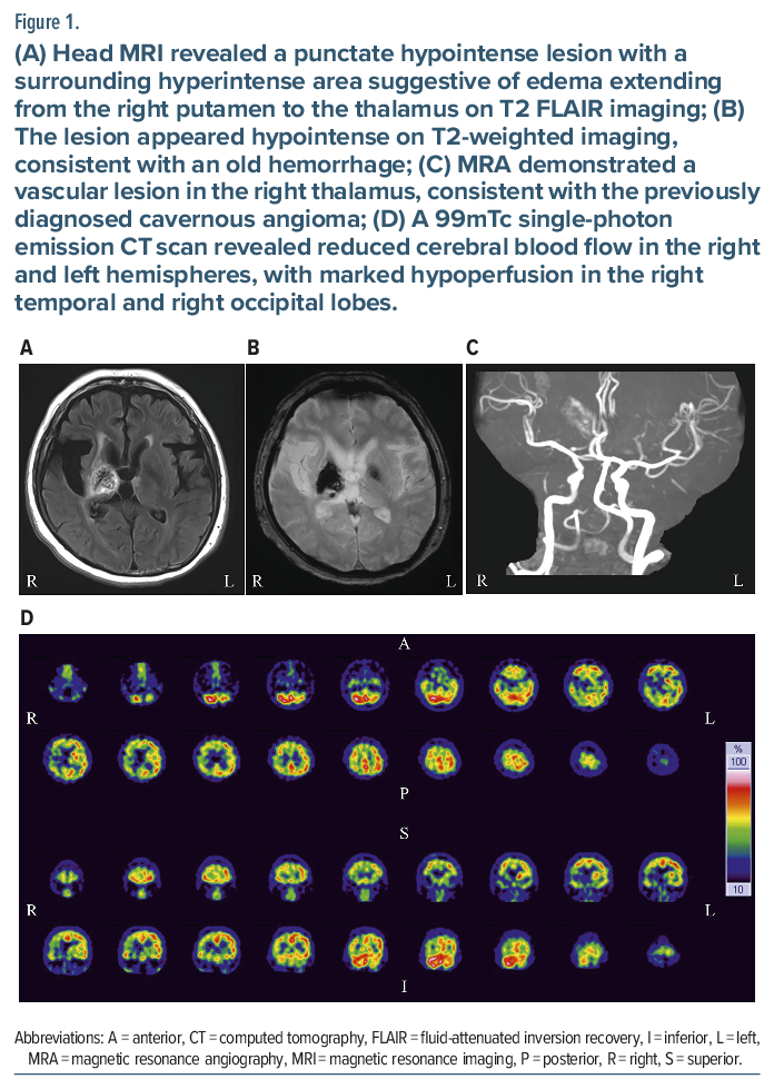
She was admitted to the psychiatry department at our hospital. Head magnetic resonance imaging revealed a punctate hypointense lesion with a surrounding hyperintense area suggestive of edema extending from the right putamen to the thalamus on T2 fluid-attenuated inversion recovery imaging (Figure 1A). The lesion appeared hypointense on T2-weighted imaging, consistent with an old hemorrhage (Figure 1B). Cerebral atrophy, particularly involving the temporal lobes, was also observed (Figure 1A). Magnetic resonance angiography demonstrated a vascular lesion in the right thalamus, consistent with the previously diagnosed cavernous angioma (Figure 1C). A 99 mTc single-photon emission computed tomography scan revealed reduced cerebral blood flow in the right (32.2 mL/100 g/min) and left (34.2 mL/100 g/min) hemispheres, with marked hypoperfusion in the right temporal and right occipital lobes (Figure 1D). Accordingly, the patient was diagnosed with peduncular hallucinosis associated with the right thalamic cavernous angioma. Aripiprazole was initiated at a dose of 6 mg/day, and this partially improved her hallucinations and delusions. The patient was subsequently discharged.
Discussion
In the present case, the right thalamic cavernous malformation was incidentally identified and initially followed an asymptomatic course. However, a hemorrhage developed gradually from the lesion, and surrounding edematous changes and alterations in the adjacent brain tissue developed. These pathological changes were considered to have contributed to the emergence of various psychiatric symptoms, especially visual and auditory hallucinations. The lateral geniculate nucleus of the thalamus serves as a relay nucleus for visual information, whereas the medial geniculate nucleus functions as a relay nucleus for auditory information; each is connected to the corresponding visual and auditory cortices via the thalamic radiations.5 In our case, impairment of the thalamic peduncle, including the optic radiation extending from the thalamus to the right occipital lobe, was suggested to be responsible for visual hallucinations, whereas impairment of the inferior thalamic peduncle, including the auditory radiation extending to the right temporal lobe, was suggested to be responsible for auditory hallucinations. These impairments may lead to peduncular hallucinosis. Further studies are needed to elucidate the functional impairments underlying peduncular hallucinosis.
Article Information
Published Online: 18 August, 2026. https://doi.org/10.4088/PCC.26cr04179
© 2026 Physicians Postgraduate Press, Inc.
Prim Care Companion CNS Disord 2026;28(4):26cr04179
Submitted: January 4, 2026; accepted March 18, 2026.
To Cite: Kudo M, Daa Y, Hirakawa H. Peduncular hallucinosis with right thalamic cavernous angioma. Prim Care Companion CNS Disord 2026;28(4):26cr04179.
Author Affiliations: Department of Neuropsychiatry, Oita University Faculty of Medicine, Oita, Japan (all authors).
Corresponding Author: Hirofumi Hirakawa, MD, PhD, Department of Neuropsychiatry, Oita University Faculty of Medicine, Idaigaoka 1-1, Hasama-machi, Yufu-city, Oita 879-5593, Japan ([email protected]).
Financial Disclosure: None.
Funding/Support: None.
Patient Consent: Consent was received from the patient to publish the case report, and information has been de-identified to protect anonymity.
ORCID: Hirofumi Hirakawa: https://orcid.org/0000-0002-7782-5807
References (5)

- Hegde AN, Mohan S, Lim CC. CNS cavernous haemangioma: "popcorn" in the brain and spinal cord. Clin Radiol. 2012;67(4):380–388.
- Van der Werf YD, Witter MP, Uylings HB, et al. Neuropsychology of infarctions in the thalamus: a review. Neuropsychologia. 2000;38(5):613–627.
- Schmahmann JD. Vascular syndromes of the thalamus. Stroke. 2003;34(9):2264–2278.
- Benke T. Peduncular hallucinosis: a syndrome of impaired reality monitoring. J Neurol. 2006;253(12):1561–1571.
- Marcuse LV, Langan M, Hof PR, et al. The thalamus: structure, function, and neurotherapeutics. Neurotherapeutics. 2025;22(2):e00550.
Please sign in or purchase this PDF for $40.